


Osteochondrosis is an outdated name for degenerative disease of the spine. The old term is often used in our country, but it does not reflect the essence of the disease, which is based on age-related degeneration - destruction of tissue structure. In this article we will consider the first signs of osteochondrosis, patterns of its development and treatment options.
What is osteochondrosis
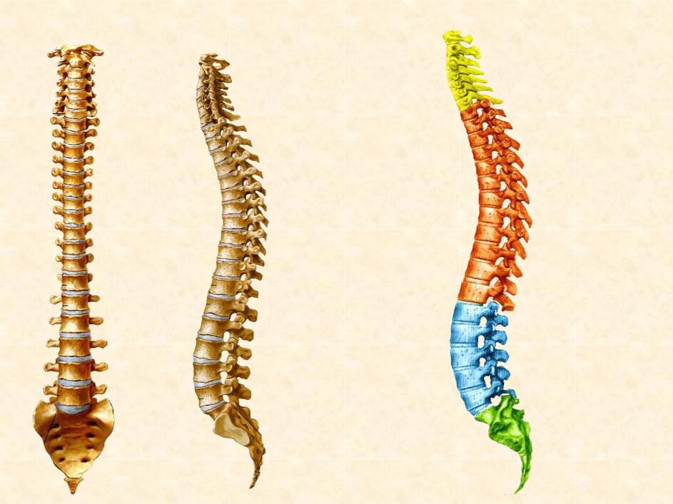
To understand the processes that occur during osteochondrosis, it is necessary to understand the anatomy of the spine. It includes the following structures:
- Vertebrae made up of bodies, arches, processes. Between the arches of adjacent vertebrae are joints called facets
- Intervertebral discs located between the bodies of adjacent vertebrae
- Spinal ligaments
- The posterior and anterior longitudinal 一 passes along the bodies of all the vertebrae in front and behind
- Ligamentum flavum: connects the arches of adjacent vertebrae
- Supraspinous ligaments and interspinous ligaments: connect the spinous processes
- The spinal cord, which is located in the spinal canal, along with the nerve roots that extend from it. They are processes of nerve cells. Through these processes, the brain receives information about the state of tissues and in response sends signals that regulate their functioning: muscle contractions, changes in the diameter of blood vessels and much more.
Degeneration begins with the intervertebral discs, and as the changes progress, all of the above-mentioned structures are involved in the process. This is partly because discs have no blood vessels. Nutrients and oxygen enter them from the vertebrae and other surrounding structures by diffusion.
The intervertebral discs make up one-third of the length of the spine and act as shock absorbers, protecting the vertebrae from overload during heavy lifting, prolonged standing or sitting, bending and twisting. Each disc consists of:
- The nucleus pulposus, which is located inside, in the center, contains a lot of hyaluronic acid, type II collagen, which retains water. This gives the normal core a gel-like consistency for effective cushioning. As degeneration progresses, the composition of the internal part of the disc changes, its water content decreases, the nucleus "dries out" and the height of the intervertebral disc decreases.
- The annulus fibrosus, which is located outside the nucleus and is made up of 15-25 layers of collagen fibers. The collagen in the annulus fibrosus is type I. It is denser than the nucleus and is necessary to hold the inside of the disc and protect it from damage. The fibers of the annulus are intertwined along the periphery with the posterior longitudinal ligament of the spine. This ensures the immobility of the spinal structures in a healthy person - doctors call this condition spinal stability. In people with degenerative disease, the fibrous ring breaks down, so instability may develop: adjacent vertebrae may move anteriorly or posteriorly relative to each other. This is dangerous due to the pinching of the nerve root between them
It is also important to mention the end plates. These are thin cartilages located between the vertebral bodies and the discs. They contain blood vessels that supply the disc. In degenerative diseases, calcium is deposited in the endplates, compromising blood supply.
Stages of development of osteochondrosis
The development of spinal osteochondrosis occurs gradually:
- Initial degeneration. The intervertebral disc does not receive sufficient nourishment, it wears out, its height decreases and it breaks. The nucleus pulposus protrudes through microdamage of the annulus fibrosus, irritating the posterior longitudinal ligament and causing pain and reflex spasm of the back muscles
- Swelling of the intervertebral disc. The fibers of the fibrous ring are destroyed, the nucleus pulposus protrudes more strongly, forming a hernia. It can compress the roots of the spinal nerves, leading to the development of paresis or paralysis of the limb muscles and a decrease in skin sensitivity. One of the complications of hernia is its sequestration: separation of the disc protrusion from its main part.
- Progression of degeneration of the protrusion and other structures of the spine. The disc becomes even more compact, and the body tries to compensate for the excessive mobility of the spine by forming pathological bone growths of the vertebral bodies - osteophytes. They, like the hernia itself, can affect nerves and ligaments, disrupting their function and causing pain. Unlike hernias, bone spurs do not dissolve.
Complications of osteochondrosis, in addition to compression of herniated spinal nerve roots:
- Spondyloarthrosis. The reduced height of the intervertebral disc subjects the facet joints to greater stress. They can develop inflammation and malnutrition, which makes them "dry" and causes pain.
- Spondylolisthesis 一 displacement of the vertebral bodies relative to each other due to ligament damage
- Degenerative processes in the area of the ligamentum flavum cause its thickening. This is dangerous because the ligamentum flavum is adjacent to the spinal canal and can narrow it, compressing the spinal cord
- At the level of the 1st-2nd lumbar vertebra, it extends from the spinal cord"ponytail" - a bundle of nerve roots responsible for the innervation of the lower extremities and pelvic organs: bladder, rectum, external genitalia. Cauda equina syndrome is one of the most dangerous complications of osteochondrosis, manifested by severe pain, muscle weakness of the legs, numbness of the perineum, urinary and fecal incontinence.
Causes of osteochondrosis of the back
There is still no consensus on what degree of degenerative changes in the spine should be considered normal. Sooner or later, the aging of the spine begins in every person.
In most people, these changes are minor and cause no symptoms: They are sometimes discovered incidentally during a magnetic resonance imaging (MRI) scan of the spine. The progression of degeneration leads to significant changes in the structure of the spine. The intervertebral discs can be so destroyed that they cease to perform the shock-absorbing function, swell and put pressure on the spinal nerves and even the spinal cord itself.
It is impossible to accurately predict how severe the degenerative changes will be in a given person and whether they will lead to complications. There is a genetic predisposition to osteochondrosis, but the specific genetic mutations responsible for the course of the disease have not been identified. Therefore, there is no accurate genetic test that can show personal risk. There are some factors that increase the risk of developing osteochondrosis. It is they who are targeted by measures to prevent osteochondrosis.
Risk factors for osteochondrosis include:
- Excessive load on the spine: professional sports, weight lifting, intense and regular physical work
- Remaining in a static and incorrect position for a long time: sitting, hunched over, cross-legged, in a chair without lumbar support, working in an upright position with an incline
- Sedentary lifestyleleading to weakness of the trunk muscles that cannot effectively support the spine
- Overweight 一 Obesity creates additional stress on the back and joints
- Smoking - Nicotine and other components of cigarettes interrupt the diffusion of nutrients from blood vessels to tissues, including intervertebral discs
- Alcohol intake - Regular consumption leads to the fact that calcium is poorly absorbed from food. The lack of calcium causes the vertebrae to lose density
- Back injuries with damage to the structure of the vertebrae or discs, due to which the recovery process occurs much slower than the degeneration process
Osteochondrosis of the spine in adults: symptoms
In the early stages of a degenerative disease, a person usually does not experience any symptoms. They occur suddenly or gradually as the disease progresses. The main manifestations are back pain and reflex spasm of the back muscles. The location of the symptoms depends on which part of the spine the problem occurs:
- Degeneration of the cervical spine leads to muscle stiffness, neck pain that radiates to the shoulder and arm or the back of the head and worsens with head movements
- Changes in the thoracic spine appear extremely rarely, since it is the most static. If a hernia occurs, pain appears between the shoulder blades
- Hernias in the lumbar region occur more often than others and are manifested by pain in the lower back or sacrum, radiating to the gluteal region, leg. Stiffness in the lower back is also noted. The pain worsens when sitting, standing for long periods of time, and bending.
If the pain radiates from the back to the limb, it is called radiculopathy: damage to the nerve root. This is compression by a herniated spinal nerve. Radiculopathy, in addition to pain, is also accompanied by other symptoms localized in a specific area innervated by the damaged nerve. Such manifestations may include:
- weakness of the limb muscles, up to paralysis
- disorders of the sensitivity of the skin of the extremities
- bladder and rectal dysfunction with lumbar radiculopathy
Signs of spinal osteochondrosis in women and men generally do not differ, but in women symptomatic degeneration develops more rapidly after menopause, when bone density decreases. In men, degenerative processes are more often caused by physical work and develop at an early age, but gradually.
Not all back pain is caused by spinal osteochondrosis. Our specialists can conduct a comprehensive examination and decide whether an MRI is necessary.
Osteochondrosis of the spine at a young age
It is generally accepted that osteochondrosis is a disease of older people. Degenerative spinal disease is indeed common among patients over the age of 60, but is becoming increasingly common in people in their 30s and 20s. Usually the cause is genetic predisposition, excess weight, sedentary lifestyle or back injuries. Both serious one-off injuries, for example due to a fall, and regular minor injuries, for example when playing professional sports, are important. The disease occurs most often in the lumbar region since it is the most mobile. Intervertebral hernias, including Schmorl's nodes, can form here. The main mechanism of their occurrence is damage to the endplates, which cannot withstand intradiscal pressure. This causes protuberances to form in the body of the upper or underlying vertebra, called Schmorl's hernias. They do not cause compression of the nerve roots and are usually not dangerous. In rare cases they can grow and cause back pain, but more often they are discovered by chance during an MRI. Intervertebral hernias that protrude posteriorly are usually accompanied by pain and may require treatment.
Osteochondrosis of the spine: treatment
Up to 90% of cases of degenerative disease can be treated conservatively.
Surgery is indicated only if serious complications threaten, such as progressive loss of bladder control or weakness of the lower limbs. Surgical treatment allows you to save a person from paralysis, but in itself does not relieve pain and further progression of the disease, therefore, after the operation, a special rehabilitation program is prescribed.
Uncomplicated hernias in many cases resolve on their own. The resorption process may be accompanied by the formation of excess connective tissue and calcifications in the spine, which increases the likelihood of disease recurrence in the future. Existing physiotherapeutic techniques and special exercises help:
- accelerate the resorption of the hernia
- improve the power of the disc
- normalize the biomechanics of movements and load distribution
- avoid the need for surgery in the future
For pain, drugs from the groups of non-steroidal anti-inflammatory drugs, glucocorticoids and muscle relaxants are also used, but the use of the drugs is limited to the acute period of the disease and does not improve the condition of the spine in the long term. It is possible to reduce the intensity of degeneration:
- MLS laser therapy - the laser radiation used has an anti-inflammatory effect, dilates the lymphatic vessels, improving lymphatic drainage
- Acupuncture: This method relieves pain, swelling and inflammation due to the body's reflex response to the stimulation of biologically active points on the body with special needles
- The magnetotherapy method stimulates blood flow, normalizing the diffusion of nutrients and removing toxins from the thickness of the intervertebral discs, accelerating recovery processes
- Therapeutic physical education: special sets of exercises help to strengthen the muscles of the torso, learn to correctly distribute the load on the back, maintain the correct posture and relieve muscle spasms. To monitor performance, it is better to start working with an instructor, and then continue the exercises yourself according to the recommendations
Depending on the manifestations of the disease and the characteristics of the patient, different combinations of the above methods can be used.
Both conservative treatment of spinal hernias and rehabilitation after surgery can be performed on an outpatient basis in the clinic. It has all the necessary equipment and a team of professionals specialized in the non-surgical treatment of hernia. It is not advisable to go to hospitals where methods without scientific basis and not approved by the world medical community are used: this can be dangerous for your health. In a modern clinic you can receive advice at an affordable price and decide on a further course of action together with your doctor.